

Newsletter 2015.11 Index
Theme : "Mechanical Engineering Congress, 2015 Japan (MECJ-15) Part 1"
|
Elucidation of the sudden death by the computational fluid dynamics
Tadashi YAMAMOTO
|

Abstract
A large part of sudden death caused by acute myocardial infarction (AMI). Based on the mechanical elucidated the evidence the onset wit of AMI, it is object that a risk of AMI onset is visualized by computational fluid dynamics (CFD). AMI is reported that a vulnerable plaque in coronary artery is ruptured by high wall shear stress (WSS) produced by coronary blood flow. WSS is calculated in the CFD for those cases that is revealed vulnerable plaque by coronary artery CT. A case in which WSS indicates a high value at vulnerable plaque, is regarded as high-risk group of AMI. To realize a life style improvement and take a proper medicine in order to reduce the WSS will prevent sudden death. CFD that has not been used in the medical field until now will be able to provide visualization of sudden death prevention.
Key words
Sudden death, Acute myocardial infarction, Atherosclerosis, Vulnerable plaque, CFD
Figures
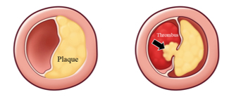
Fig. 1 Image of plaque rupture Arrow: rupture point

Fig. 2ツImage of plaque rupture due to high WSS
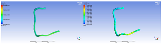
Fig. 3ツ Image of stream line and WSS in normal coronary artery
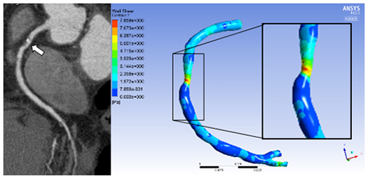
Fig. 4 (Left) Image of coronary artery CT. Arrow: vulnerable plaque.
(Right) Image of WSS; Red indicate high WSS