

Newsletter 2026.3 Index
Theme : "The Eleventh JSME-KSME Thermal and Fluids Engineering Conference (TFEC11) "
|
Electrical Tomography × Flow Visualization × Startups
– Chiba University Spin-off Startups and the Future of Fluid Engineering –
 |
 |
Songshi LI
|
Masahiro TAKEI Chiba University |
Abstract
In this article, we will provide a statistical summary of the current state of university-based startups, introduce Chiba University's entrepreneurship education initiatives, and introduce a startup founded by our laboratory with the aim of achieving a future exit. In the summer of 2025, the founding of TOMOCLOUD marked the launch of a startup spun out of Chiba University. Leveraging electrical impedance tomography (EIT), a core technology of the Takei Laboratory, visualization measurement equipment is being developed to reconstruct images of the internal state of living organisms and fluidic devices in real time. EIT is a completely different approach from conventional X-ray CT and MRI, enabling 3D visualization and measurement. By applying weak electrical currents, EIT allows non-invasive, radiation-free monitoring of biological tissues, offering the potential for safe, continuous observation and early disease detection. Through this case study, this article discusses the prospects of university-based startups in the field of fluid engineering and bioengineering, and highlights the role of advanced measurement technologies in bridging academic research and practical societal applications.
Key words
Fluids visualization, Electrical impedance tomography (EIT), Startup
Figures
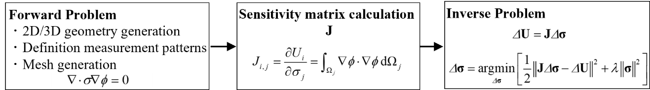
Fig. 1 Image reconstruction flow of ET
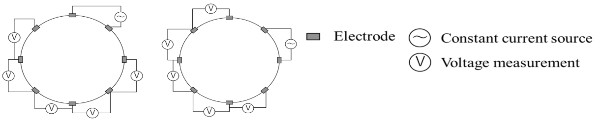
Fig. 2 Electrode pattern
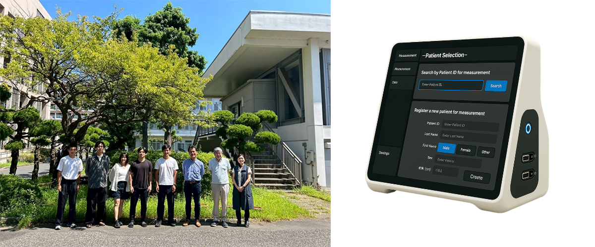
Fig. 3 Chiba University startup members and LT monitor
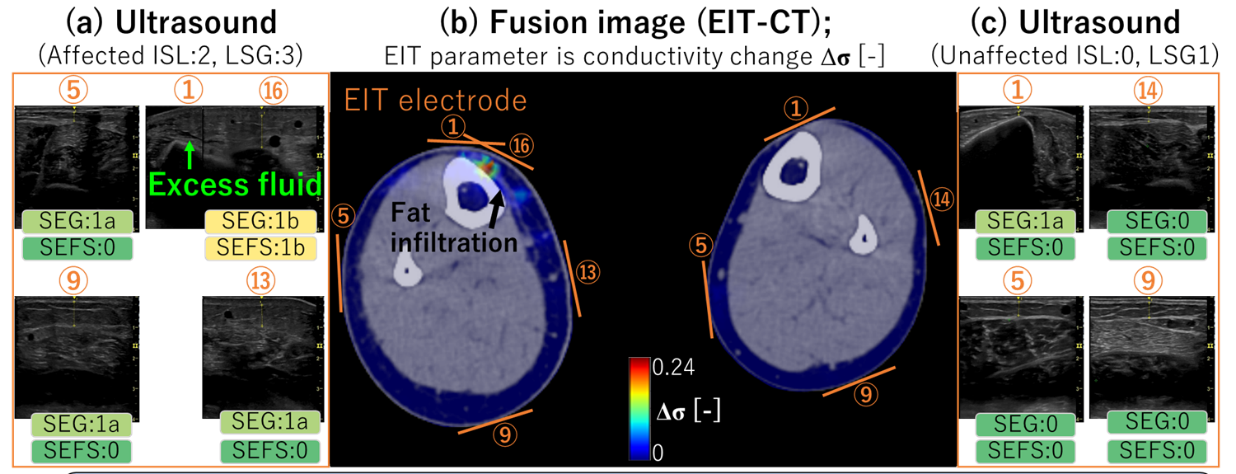
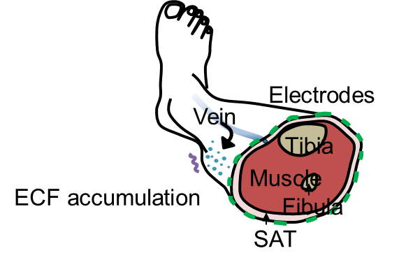
High electrical conductivity is observed near the great saphenous vein (GSV).
CT images show fat infiltration, while ultrasound images reveal excessive interstitial fluid.
These findings suggest lymphatic fluid stagnation with backflow from the dermis.
Fig. 4 Clinical application of the LT monitor and comparison with X-ray CT and ultrasound images